Neuromuscular Ultrasound
What is Neuromuscular Ultrasound?
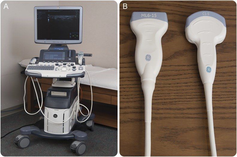
Neuromuscular Ultrasound is a diagnostic tool for nerve and muscle diseases that provides real-time anatomic and physiologic information.
A Neuromuscular Ultrasound scan is a high-resolution ultrasound to image the peripheral nervous system of patients with suspected neuromuscular diseases, myopathies, neuropathies, and motor neuron disorders.
What Nerve Disorders Can Be Diagnosed?
Neurologists use neuromuscular ultrasound as an extension of diagnostics for peripheral nerve disorders, including

- Hereditary neuropathy
- Nerve trauma
- Entrapment neuropathy (well, which is a nerve trauma)
- Infection, especially leprosy
- Auto-immune disease
- Nerve tumours
When is Neuromuscular Ultrasound Used?
It is typically used with standard diagnostic tests such as
- electromyography,
- nerve conduction studies, and
- tissue biopsy.
Neuromuscular Ultrasound allows for studying muscle contraction, fasciculations, and nerve and tendon movement. This form of testing can complement peripheral nerve assessments (nerve conduction studies, electromyography) to understand:
Peripheral Nerves ultrasound:
- Nerve enlargement
- Quantification of the swelling
Muscles Ultrasound
- Changes in thickness,
- Echogenicity and
- The architecture of the muscle, and
- Documenting involuntary movements (eg. in motor neuron diseases)
What is Different from Normal Ultrasound?
Neuromuscular Ultrasound differs from the classical use of ultrasound in its focus on the “neuromuscular system” this means focusing on the visualisation of nerves and muscles to aid the diagnosis of either.
- Mono- and polyneuropathies or
- Myopathies
of different origin. When searching for neuropathies, also alterations of the muscles supplied by a specific nerve as a guide.
The visualisation of nerves and muscles can be used as help for applying targeted therapy and performing optimised electromyographic studies.
As a sensitive and specific tool for detecting many neuromuscular disorders, it can also be used as a screening tool among children with suspected peripheral nervous system diseases.
Who can Benefit from Neuromuscular Ultrasound?
Depending on which nerves need to be assessed, a Neuromuscular Ultrasound may need to deal with some really small structures of a diameter less than 1 mm.
This allows for your doctor to isolate not only the right muscle but also to examine the pathology.
Preparation for a Breast Ultrasound
What Special Diet is required Before an Ultrasound?
There are no special diets required before an ultrasound. However, you may be instructed to drink extra fluids or abstain from eating, depending on the type of ultrasound procedure.
What should a Patient Tell the Radiographer Before an Ultrasound?
Before undergoing an external ultrasound to examine your blood vessels and blood flow, you should tell the radiographer if you are diabetic.
What to bring or wear for an ultrasound?
You don’t need to bring anything with you. Everything needed will be supplied by the radiograph.
The are no special clothing requirements, although you will be required to remove your top for a Breast Ultrasound, so you should try and wear something comfortable and easy to remove.
You will be provided with a place to change and a gown to wear.
How Long will the Ultrasound Take?
Depending on the type of ultrasound and the part of the body being examined, the procedure can take anywhere from 15 to 45 minutes.
Ultrasound Procedure Description
What does Breast Ultrasound Involve?
Ultrasounds are usually administered in hospitals or specialist medical centres by trained staff, such as radiographers, sonographers, midwives or physiotherapists.
The procedure itself is very simple, although the exact procedure will vary depending on what type of ultrasound is needed.
Specific steps during an Ultrasound
The ultrasound procedure varies depending on which body part is being examined. For example:
- During an external Ultrasound, a lubricating gel is placed on the skin, and then the handheld probe is passed over the area.
- During an internal Ultrasound, you will need to lie down on your back or your side with your legs bent. A small probe is then inserted into the cervix or rectum.
- During an endoscopic Ultrasound, the probe is attached to a long narrow tube. Typically you will be given a sedative, and a local anesthetic spray will be used to numb your throat. The probe is then inserted into the mouth and fed down the throat.
Your GP and radiographer will explain the procedure to you and answer any questions you may have.
Post Ultrasound Instructions
Side effects are rare; you will be able to return to work or home immediately after an ultrasound.
Patients who receive an external on internal ultrasound will be able to drive immediately after the procedure.
Get in Touch
Once your referral is triaged, an appointment date and time will be made for you.
About Us
Neuro NSW provide multi disciplinary care and are a multi specialist practice. We provide extensive Neuro diagnostic testing and comprehensive Neurological assessment.
Get in Touch
- Mon - Thu
- -
- Fri - Sun
- Closed
- Public Holidays: Closed
- Closed from 22/12/2023 - 03/01/2024
- Emergency: Not
available
All Rights Reserved | Neuro NSW Brain & Spine Specialists


